Every thought and action is carried out by cells that signal to one another. This unit explains how a neuron fires and how it passes its message across a synapse, what drugs such as caffeine and nicotine do there, how the nervous system and the brain are organized, and what lesions from Broca's patient to Phineas Gage showed about which region does what. It ends with the adult brain's capacity to change and with the first real count of its cells.
Preview: the beginning of the unit
Every thought, reflex, and memory described in this masterclass is carried out by cells that signal to one another. This unit is about those cells and the organ they build. It starts small, with the electrical pulse that one neuron sends along its axon and the chemical message it hands to the next cell, and it ends large, with the regions of the brain and what each of them does. On the way it meets a railway foreman who survived an iron rod through his skull, a patient who could say only one syllable, and a laboratory that counted the cells of the human brain and found that the textbooks had been wrong for decades.
1 The neuron and its signal
1.1 The cells of the nervous system
Nervous tissue is made of two kinds of cells. The neuron is the cell that carries signals, and the glia are the cells that support it, insulate it, feed it, and clean up after it. For a long time nobody knew whether neurons were separate cells at all, and the question was settled by two men who could not agree about it.
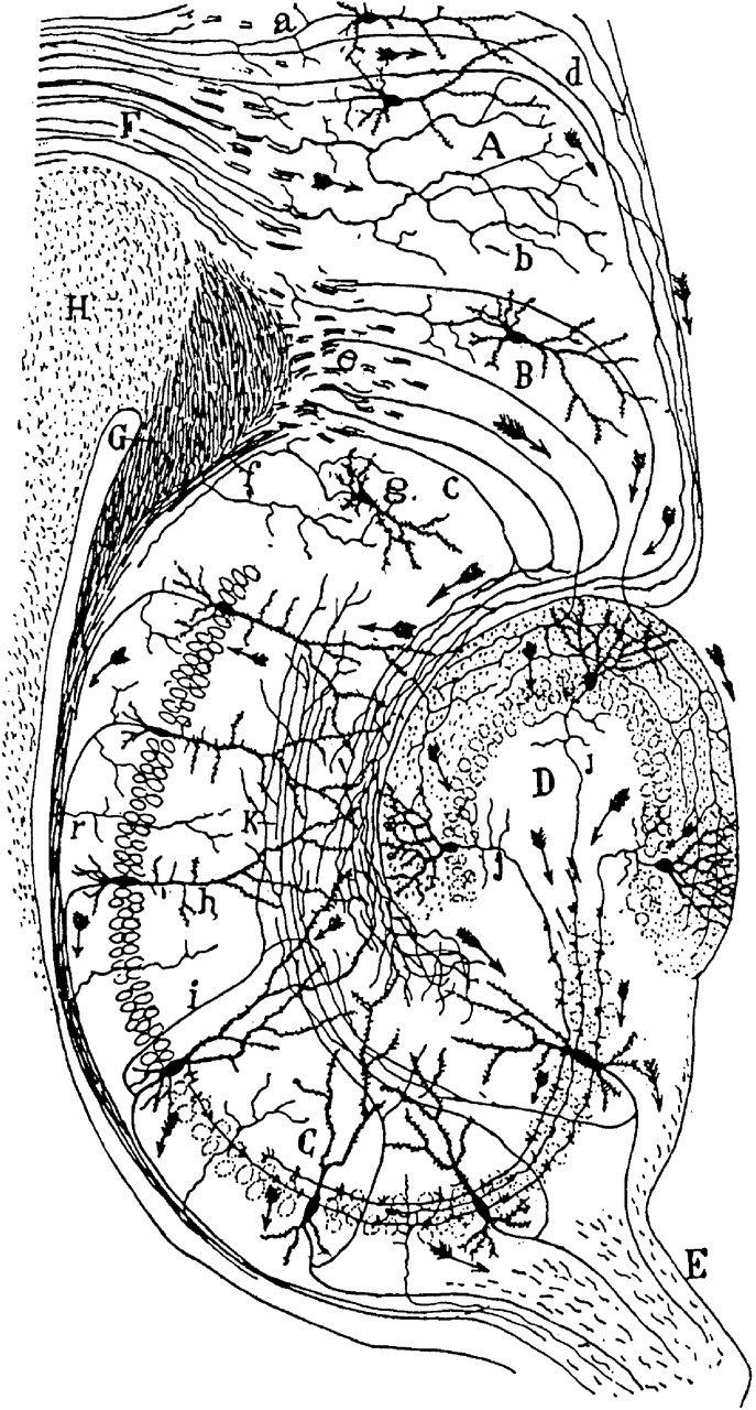
Figure 1. Cajal’s drawing of the hippocampus of a rodent, from his textbook of 1911, stained by Golgi’s method. The arrows mark the direction in which he inferred that the signals travel from cell to cell. Credit: Santiago Ramón y Cajal, public domain, via Wikimedia Commons.
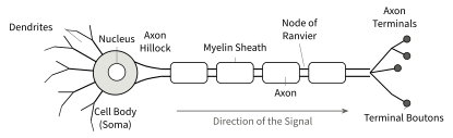
Figure 2 shows the parts of a typical neuron. The dendrites are branched extensions that receive signals from other cells. The cell body, also called the soma, holds the nucleus and keeps the cell alive. The axon is a single thin fibre that carries the cell’s signal away from the cell body, over a distance that ranges from less than a millimetre in the cortex to more than a metre in the nerve that runs from the spinal cord to the toes. It leaves the cell body at a small cone, the axon hillock, which is where the signal starts. Near its end the axon branches, and each branch ends in a swelling called a terminal bouton (bouton is the French word for button), which sits close to the next cell. Many axons are wrapped in myelin, a fatty insulating sheath laid down by glial cells, with short gaps between the segments called nodes of Ranvier. Section 1.3 explains why the gaps matter.
Figure 2. The parts of a neuron. Signals arrive at the dendrites and the cell body, start at the axon hillock, run along the myelinated axon, and reach the terminal boutons, which contact the next cells.
The glia come in four main kinds.
Astrocytes feed neurons, control the chemistry of the fluid around them, and form part of the blood-brain barrier, the tight lining of the brain’s blood vessels that lets few substances through.
Oligodendrocytes make the myelin inside the brain and spinal cord.
Schwann cells make the myelin in the nerves of the body.
Microglia are the immune cells of the brain.
Textbooks used to say that glia outnumber neurons ten to one. They do not, and Section 7 tells how that was found out.
The full unit is part of the program
Want to see a complete unit first? Unit 3, Sensation and Perception, is free to read.