

Memory in the Brain
What Henry Molaison's brain showed when it was sectioned, how a synapse is strengthened by long-term potentiation and why the NMDA receptor detects coincidences, how place cells and grid cells map the space around an animal, how sleep moves a memory into the cortex, and how a memory was tagged and switched on with light in mice. The unit ends with Alzheimer's disease and with what the brain evidence adds to the testing and spacing effects.
Unit 5 described memory from the outside, as what people and patients can and cannot recall. This unit opens the skull. It follows a memory from the synapses that change when something is learned, through the cells of the hippocampus that map the space around an animal, to the slow work of sleep that moves a memory into the cortex, and finally to the experiments in which a memory was switched on with light. The patient with whom Unit 5 began, Henry Molaison, returns at the start, because the examination of his brain after his death in 2008 showed more precisely than any scan what his operation had taken and what it had left.
1 The medial temporal lobe
On 1 September 1953 the neurosurgeon William Beecher Scoville (1906-1984) removed the inner part of both temporal lobes of a 27-year-old patient, and from that day on patient H.M., Henry Molaison (1926-2008), could no longer form lasting memories of events and facts (Scoville and Milner 1957). Which of the removed tissues made the difference was settled only after his death. In December 2009 a team led by Jacopo Annese cut his brain into 2,401 slices of 70 micrometres, each about as thick as a human hair, over 53 hours, photographed every cut surface, and built a digital three-dimensional model from the images (Annese et al. 2014). Magnetic resonance scans had already shown that the removal reached about 5.4 cm back from the tip of the left temporal lobe and 5.1 cm on the right (Corkin et al. 1997); the sections confirmed the left figure, shortened the right one to 4.4 cm, and added the details. The amygdala was almost entirely gone. The entorhinal cortex, the sheet of cortex through which the hippocampus talks to the rest of the brain, was reduced to about 1.7% of its normal volume on the left and 6.5% on the right. About half of the hippocampus had been removed, and the half that remained at the back, roughly 24 mm of it on each side, had lost the entorhinal cortex that supplied its input, so it was tissue without a working connection. The parahippocampal cortex further back was largely intact, and the cerebellum was shrunken, which the authors attributed to the anti-epileptic drug phenytoin that he had taken for decades, because the operation had been an attempt to treat his epilepsy and had not removed the need for the drug.
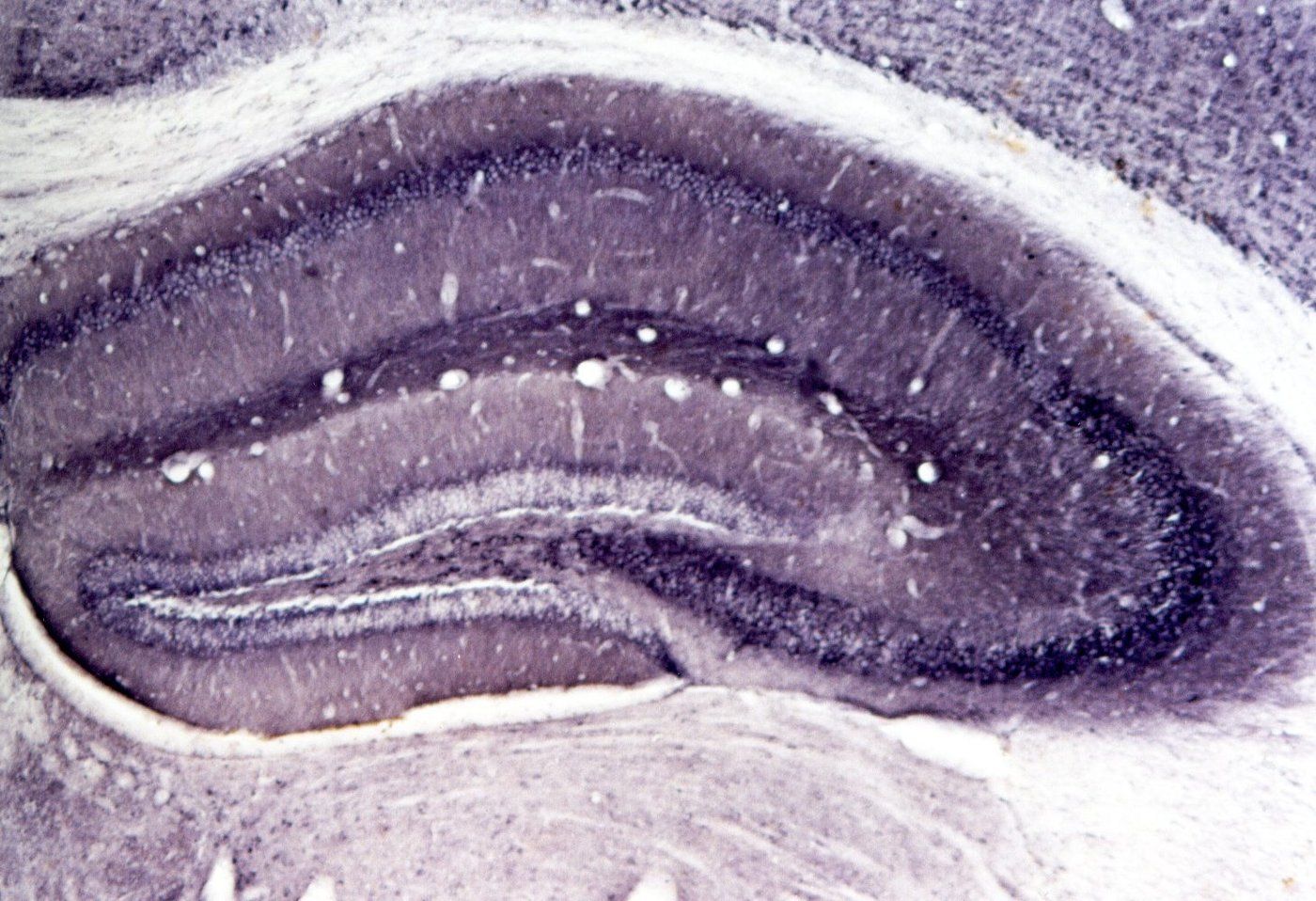
The anatomy that this examination mapped is drawn in Figure 1. The hippocampal formation is a curled strip of cortex of an evolutionarily old and simple type along the inner edge of the temporal lobe, and in section it looks like two interlocking C shapes; Figure 2 shows the two in a real section. One C is the dentate gyrus, a dense band of small granule cells. The other is the hippocampus proper, divided into the fields CA3 and CA1 (CA stands for cornu ammonis, the ram’s horn), and it continues into the subiculum, which leads back out to the entorhinal cortex. Next to the entorhinal cortex on the underside of the temporal lobe lie the perirhinal cortex, on the two banks of a groove called the collateral sulcus, and the parahippocampal cortex behind it.
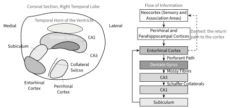
Information flows through this system in a loop (Figure 1, right). Sensory and association areas of the neocortex send their signals to the perirhinal cortex, which deals mostly with objects, and to the parahippocampal cortex, which deals mostly with scenes. Both project to the entorhinal cortex, which is to say that they send their fibres there. From there the perforant path enters the dentate gyrus (with direct branches to CA3 and CA1 as well), the granule cells send mossy fibres to CA3, and CA3 sends the Schaffer collaterals to CA1; this chain of three synapses is known as the trisynaptic circuit. CA1 projects to the subiculum and back to the entorhinal cortex, and from there the signal returns to the neocortical areas it came from. The loop is the reason the hippocampus can bind together the sight, the sound, the place, and the time of one event that are stored in different parts of the cortex, and the reason a lesion in it leaves those cortical areas intact while removing the binding.
Henry Molaison’s operation showed a second thing, which Unit 5 reported: his memory for events and facts failed, but he learned mirror drawing (tracing a shape that he could see only in a mirror), he improved at puzzles he did not remember having seen, and he could keep a number in mind as long as he was not distracted. Each spared ability has since been traced to tissue that the surgery had not touched (Table 1), and the rest of this unit is about the one system, the declarative one, whose core the operation had removed.